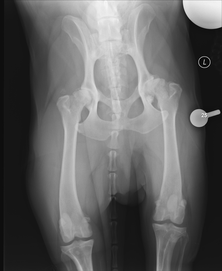
In my faculty appointment at the Cummings Schools of veterinary medicine at Tufts University here in Grafton MA, I have been working with the surgeon who runs the clinical skills simulation center at Grafton. We decided a cool project to work on is a series of 3D printed training models around hip-dysplasia (a fairly common problem in pure bred dogs such as german Shepards, and not super rare in human babies either). When my wife heard about the project (she is a graduate from Tufts Vet) she suggested it would be more helpful to have the x-ray showing behind the model so students, owners and community vets can learn to associate the x-ray findings with the actual models. So I got some of the x-rays for dogs with the condition, and planned an edge-lit version.
Moderate Hip Dysplasia
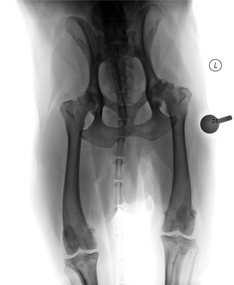
And at first could not get a successful engrave to happen (thanks to @Jules) we figured out that what appears black on the x-ray (i.e. the background) is of course not actually black to the GFUI (it’s very, very close but #010101 is not #000000 to software. So a quick trip to photoshop and playing with the levels slider, then inverting the image:
(the lollipop thing is a scale device so you can judge exact distances on the x-ray). I also realized that to make this thing way cooler in a lit room you need to make the background black so I cut a second piece of black acrylic to glue on the back (of course to be able to insert the whole thing in the lit base I chopped the tab at the bottom that inserts into the light). Then using Weldon for acrylic a drop in teach corner pulled in by capillary action welded the black on
Then off to the GF to engrave:
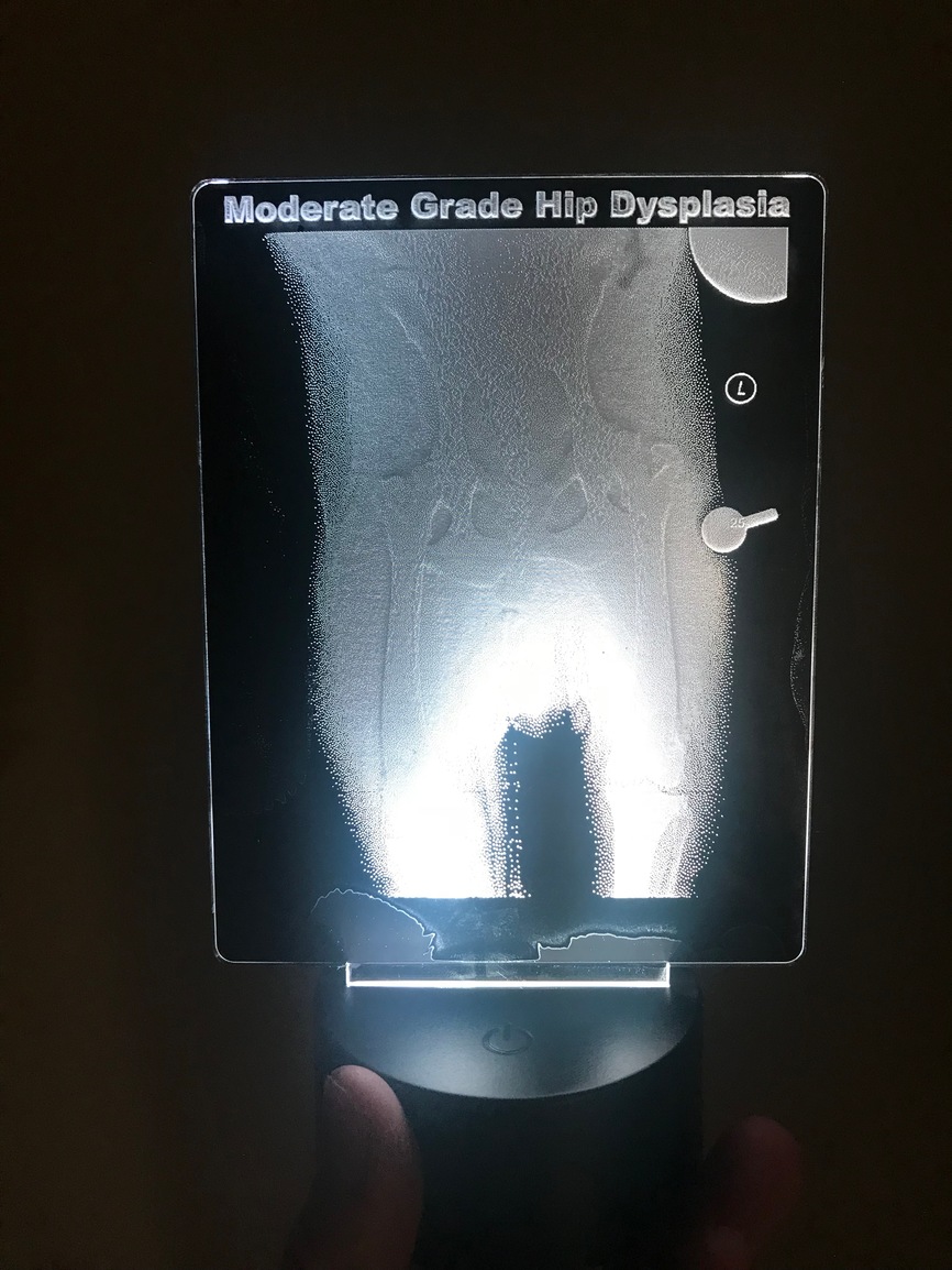
I didn’t come out as nicely as I hoped and will be working on the image to see if I can improve it, but it is a fairly cool effect (and the iPhone can’t really photograph it well). The bones appear to be floating above the image. I think I need to make the bones way higher contrast and make the soft tissues way, way less visible. Anyways, figured I’s share so folks can learn from this.
